Call Us : 01233 661662
- About Us
- Associated Clinics
- Treatment
- Line & Wrinkle Freezing
- Wrinkle Fillers
- Facial Implants
- Skin Peels
- Active skin care
- Acne treatment
- Mole, Skin and Wart Removal
- Pigmented Lesions
- Leg and Facial Veins
- Microdermabrasion
- Stretch mark removal
- Thread vein removal
- Hair removal
- Tattoo removal
- Mesotherapy
- Laser Skin Therapy
- Laser Fungal Nail Infection
- Skin Regeneration
- Sublative Rejuvenation
- Skin Tightening
- Excessive Sweating
- Polycystic OS
- Aqualyx
- PRP Platelet-Rich Plasma
- Syringoma
- Contact
What is Onychomycosis?
Fungal infection of the nails is known as onychomycosis
. It is increasingly common with increased age. It rarely affects children.
Responsible organisms
Onychomycosis can be due to:
- Dermatophytes such as Trichophyton rubrum (T rubrum), T. interdigitale. The infection is also known as tinea unguium.
- Yeasts such as Candida albicans.
- Moulds especially Scopulariopsis brevicaulis and Fusarium species.
Clinical features
Onychomycosis may affect one or more toenails and/or fingernails and most often involves the great toenail or the little toenail. It can present in one or several different patterns:
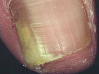
- Lateral onychomycosis. A white or yellow opaque streak appears at one side of the nail.
- Subungual hyperkeratosis. Scaling occurs under the nail.
- Distal onycholysis. The end of the nail lifts up. The free edge often crumbles.
- Superficial white onychomycosis. Flaky white patches and pits appear on the top of the nail plate.
- Proximal onychomycosis. Yellow spots appear in the half-moon (lunula).
- Complete destruction of the nail.
Tinea unguium often results from untreated tinea pedis (feet) or tinea manuum (hand). It may follow an injury to the nail.
Candida infection of the nail plate generally results from paronychia and starts near the nail fold (the cuticle). The nail fold is swollen and red, lifted off the nail plate. White, yellow, green or black marks appear on the nearby nail and spread. The nail may lift off its bed and is tender if you press on it.
Mould infections are usually indistinguishable from tinea unguium.
Onychomycosis must be distinguished from other nail disorders such as:
- Bacterial infection especially Pseudomonas aeruginosa, which turns the nail black or green.
- Psoriasis.
- Eczema or dermatitis.
- Lichen planus.
- Viral warts.
- Onycholysis
- Onychogryphosis (nail thickening and scaling under the nail), common in the elderly.
Nail clippings
Clippings should be taken from crumbling tissue at the end of the infected nail. The discoloured surface of the nails can be scraped off. The debris can be scooped out from under the nail.
Previous treatment can reduce the chance of growing the fungus successfully in culture so it is best to take the clippings before any treatment is commenced:
- To confirm the diagnosis - antifungal treatment will not be successful if there is another explanation for the nail condition.
- To identify the responsible organism. Moulds and yeasts may require different treatment from dermatophyte fungi.
- Treatment may be required for a prolonged period and is expensive. Partially treated infection may be impossible to prove for many months as antifungal drugs can be detected even a year later.
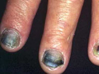
Onychomycosis from T rubrum
Complete nail destruction
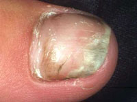
Lateral Onychomycosis
Nail infection due to Microsporum canis (rare)
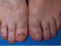
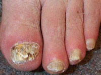
All nails are yellow due to T rubrum infection
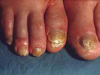
Extensive tinea unguium due to T rubrum
Treatment
Fingernail infections are usually cured more quickly and effectively than toenail infections.
Mild infections affecting less than 80% of one or two nails may respond to topical antifungal medications but cure usually requires an oral antifungal medication for several months. Combined topical and oral treatment is probably the most effective regime.
Recently, laser treatment has become available in New Zealand and elsewhere to treat onychomycosis unsuitable for or resistant to antifungal medications. There are at least three types of toenail laser, all emitting infrared radiation. Laser treatment is reported to safely eradicate nail fungi with one to three, almost painless, sessions. However, laser treatment for fungal nail infection is not yet FDA approved and it is as yet unclear how well it works.